Authors: Lauren Shin, MD; Angela M. Curcio, MD
Floating Hospital at Tufts Medical Center, Boston, MA
Reviewed: April 2022
SUMMARY
Craniosynostosis occurs when one or more of the bones of a baby’s skull fuse too early. It is mostly seen by itself, but it can be a symptom of a bigger disease. If it is not treated, it can cause serious complications.
When a baby is born, the skull has multiple bone pieces. As the baby grows, these bones join together to form the skull as we know it. The gap between the bone pieces are called “sutures.” These gaps are filled with flexible materials. This flexibility of the skull at birth:
- Allows the baby to be born through a birth canal
- Allows the baby’s brain to grow bigger as it matures
A baby’s sutures usually close over time. If the bones come together too early, the growth of the brain may be slowed or stopped.
JUMP TO
Disorder Overview
DESCRIPTION
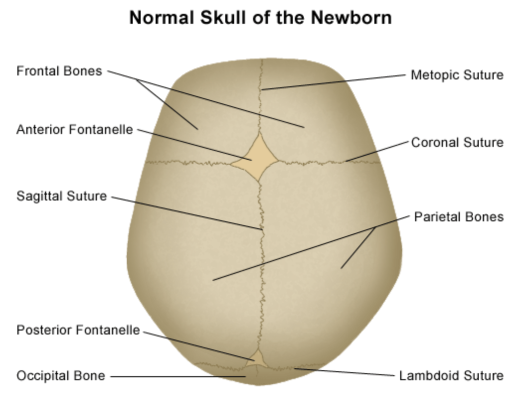
There are 4 major types of sutures of the skull. Any of these sutures can fuse too early and cause craniosynostosis. The type of craniosynostosis is named after the suture that closes too soon.
Image from Stanford Children’s Health
- Metopic suture: This suture runs in middle of the forehead, from the nose to the top of the head. If this suture closes early, the baby’s forehead may look triangular.
- Coronal suture: The left and right coronal sutures run over the top of the head between left and right ears. If one or both sides close early, the baby’s forehead will look flattened.
- Sagittal suture: This suture runs at the top of the head, from the baby’s soft spot (the anterior fontanelle) to the back of the head. If this suture closes early, the baby’s head will be long and narrow. It is the most common type of craniosynostosis.
- Lambdoid suture: The left and right lambdoid sutures run behind the head between the left and right side of the back of the head. It meets the anterior fontanelle at the back of the head. If one side or both sides close early, the baby’s head may look flat in the back.
SIGNS AND SYMPTOMS
An early fusion of the skull bones can result in:
- A misshapen head
- A small head size
- Developmental delays
- Vision and hearing impairment
- Dental abnormalities
- Breathing problems
- Decreased IQ
- Psychological impairment
- Increased pressure in the skull
Symptoms of Increased Pressure in the Skull
Sometimes a baby with this condition has symptoms of increased pressure in the skull. This is due to a lack of space for the brain and the fluid around the brain.
Symptoms of increased pressure can look like:
- Vomiting
- Poor feeding
- Lethargy
- Irritability
- Seizures
- Bulging eyes
- Not meeting developmental milestones
CAUSES
It is not clear why this disorder occurs. Craniosynostosis can appear in otherwise healthy babies. Sometimes, the baby has other problems in addition to the craniosynostosis. If other abnormalities are found, further investigations may be needed to diagnosis the underlying medical condition. Some examples of underlying causes include:
Genetic differences.
Differences during pregnancy.


LABORATORY INVESTIGATIONS
Early fusion of the skull can sometimes be seen on a prenatal ultrasound during the pregnancy. However, most of the time, it is noticed in the first 6 months of life.
Signs in the first 6 months after birth can include:
- A head shape that is not normal
- A fontanelle not felt by the pediatrician
The head may appear too long, too wide, too small, or asymmetric. Sometimes, the baby will have a raised edge over the part of the skull where the sutures are located; this is called “ridging.”
A pediatrician will refer a baby to specialists if craniosynostosis is a concern. A thorough physical examination and measurement of skull dimension can reveal the area of the early fusion. A specialist may need further investigations to look at the bones more closely. These can include:
- A skull X-ray
- An ultrasound
- A three-dimensional computed tomography scan (CT scan)


TREATMENT
If craniosynostosis is diagnosed, a neurosurgeon may perform surgery to create more space for the brain to grow. This can help with development. There are two main surgical approaches:
Separating the fused bone.
Remodeling the skull.
Babies with mild craniosynostosis may not need surgery.
OUTLOOK
Most children have a healthy life after treatment. A child’s pediatrician and specialist will continue to follow up after the surgery to make sure the baby is developing well.
The baby may need early intervention services to help with developmental delays.
Resources
Children’s Craniofacial Association
The mission of Children’s Craniofacial Association (CCA) is to empower and give hope to individuals and families affected by facial differences. Nationally and internationally, CCA offers financial assistance for medical travel, free books and educational curriculum for schools, and webinars on YouTube. Family programs and services include networking, newsletters, annual retreat, and public awareness.
The mission of Cranio Care Bears is to spread awareness, support, and compassion through loving care packages to families of children facing surgery for craniosynostosis. Our care packages include items for the child and family to relieve the stress accompanying this very serious surgery.


Child Neurology Foundation (CNF) solicits resources from the community to be included on this webpage through an application process. CNF reserves the right to remove entities at any time if information is deemed inappropriate or inconsistent with the mission, vision, and values of CNF.
Research
ClinicalTrials.gov for Craniosynostosis (birth to 17 years)
These are clinical trials that are recruiting or will be recruiting. Updates are made daily, so you are encouraged to check back frequently.
ClinicalTrials.gov is a database of privately and publicly funded clinical studies conducted around the world. This is a resource provided by the U.S. National Library of Medicine (NLM), which is an institute within the National Institutes of Health (NIH). Listing a study does not mean it has been evaluated by the U.S. Federal Government. Please read the NLM disclaimer for details.
Before participating in a study, you are encouraged to talk to your health care provider and learn about the risks and potential benefits.
Family Stories
The Children’s Craniofacial Association has been existence for over 30 years. Their Blog page shares 30 stories and 30 faces in honor of the families they have supported over the years.
On the Cranio Care Bears website, read the success stories of many children with Craniosynostosis. Lovingly shared by families and grouped by type of Craniosynostosis.
The information in the CNF Child Neurology Disorder Directory is not intended to provide diagnosis, treatment, or medical advice and should not be considered a substitute for advice from a healthcare professional. Content provided is for informational purposes only. CNF is not responsible for actions taken based on the information included on this webpage. Please consult with a physician or other healthcare professional regarding any medical or health related diagnosis or treatment options.
References
Facts about craniosynostosis [Internet]. Centers for Disease Control and Prevention. Centers for Disease Control and Prevention; 2020 [cited 2022 Mar 21]. Available from: https://www.cdc.gov/ncbddd/birthdefects/craniosynostosis.html
Mathijssen IMJ; Working Group Guideline Craniosynostosis. Updated guideline on treatment and management of craniosynostosis. J Craniofac Surg. 2021 Jan-Feb 01;32(1):371-450. https://doi.org/10.1097/SCS.0000000000007035. PMID: 33156164; PMCID: PMC7769187.
Thank you to our 2023 Disorder Directory partners:


